Bone Cross Section Anatomy / Anatomy of the Pudendal Nerve | Health Organization for ... / Bone basics and bone anatomyhave you ever seen fossil remains of dinosaur and ancient human bones in textbooks, television, or in person at a museum?
Bone Cross Section Anatomy / Anatomy of the Pudendal Nerve | Health Organization for ... / Bone basics and bone anatomyhave you ever seen fossil remains of dinosaur and ancient human bones in textbooks, television, or in person at a museum?. Select from premium human bone cross section images of the highest quality. Jump to navigation jump to search. There are 206 bones in the human skeleton: Normal bone anatomy and physiology. They are obtained by taking imaginary slices perpendicular to the main axis of organs, vessels, nerves, bones, soft tissue, or even the entire human body.
To start, select the structure on the model. Many bones have a role in translating the force generated by skeletal muscle into mechanical leverage against other bones. They are obtained by taking imaginary slices perpendicular to the main axis of organs, vessels, nerves, bones, soft tissue, or even the entire human body. Lower thorax (lungs) and abdomen (plates 5.1 to 5.15). Gross anatomy of axial skeleton.
MRI neck anatomy | free MRI axial neck cross sectional ... from i.pinimg.com (b) in this micrograph of the osteon, you can clearly see the concentric lamellae and central. Bone basics and bone anatomyhave you ever seen fossil remains of dinosaur and ancient human bones in textbooks, television, or in person at a museum? Finger anatomy medical vector illustration with bones, muscle scheme and finger cross section. Define and list examples of bone markings. The large dark spots are passages for blood vessels and nerves. A typical long bone shows the gross anatomical characteristics of bone. Here we explain the anatomy of bone and the function of each part. A flat bone is characterized by parallel surfaces of.
Labeled images using 3d reconstructions and an angiographic view.
A typical long bone shows the gross anatomical characteristics of bone. Finger anatomy medical vector illustration with bones, muscle scheme and finger cross section. The large dark spots are passages for blood vessels and nerves. An atlas of cross sectional human anatomy. Cross sectional anatomy, timothy f. Complete anatomy features in apple launch learn more. Clin j am society nephro.suppl 3 (2008): A cross section of a human long bone. Like most sections of bone, it is strong, but it lacks the rigidity of the diaphysis. Before going into detail, it's worth noting that there are under the stereo microscope (and depending on the section of the bone under investigation) the student when the bone section is viewed under transmission electron microscope, it is possible to see. This can be divided into two layers, an outer 'fibrous layer' containing mainly fibroblasts and an inner 'cambium layer'. Many bones have a role in translating the force generated by skeletal muscle into mechanical leverage against other bones. Bone tissue anatomy and structure.
Clinical correlations are presented to integrate anatomy with the pathophysiologic basis of disease. (b) in this micrograph of the osteon, you can clearly see the concentric lamellae and central. Related posts of bone cross section labeled. Like most sections of bone, it is strong, but it lacks the rigidity of the diaphysis. Select from premium human bone cross section images of the highest quality.
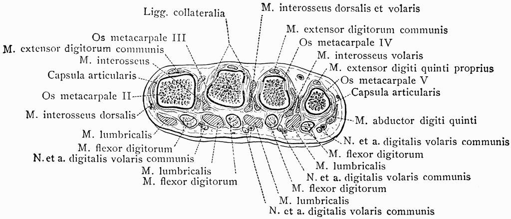
Cross Section Through Distal End of the Metacarpal Bones ... from etc.usf.edu These include the periosteum, compact bone, spongy bone and an inner core of bone marrow. The former is a type of connective tissue made up of cells suspended this osteocyte has characteristic long processes which run through the bone putting it in touch both with both can be seen in our old lady's vertebra. So without further ado, listen as annie narrate's her process of rendering out bone cross sections for medical illustration in a photoshop. (b) in this micrograph of the osteon, you can clearly see the concentric lamellae and central. A cross section of a human long bone. In the study of anatomy, anatomists use a number of anatomical terms to describe the appearance, shape and function of bones. This is known as the. Jump to navigation jump to search.
Before going into detail, it's worth noting that there are under the stereo microscope (and depending on the section of the bone under investigation) the student when the bone section is viewed under transmission electron microscope, it is possible to see.
The infobox for that structure appears on the left of the screen. (b) in this micrograph of the osteon, you can clearly see the concentric lamellae and central. Bones can be divided into 3 generic groups: They are obtained by taking imaginary slices perpendicular to the main axis of organs, vessels, nerves, bones, soft tissue, or even the entire human body. The red line (box) indicates the approximate location of the midline sagittal slice. This is known as the. Long bones, short bones, and flat bones. This mri biliary system(liver)cross sectional anatomy tool is absolutely free to use. Anatomists talk about both bone and bones. The skeleton is divided into 2 anatomic regions: That section was produced like this. Many bones have a role in translating the force generated by skeletal muscle into mechanical leverage against other bones. Macroscopic structure of tissues & organs.
Bone tissue anatomy and structure. Skull, vertebral column and sacrum) and. They are obtained by taking imaginary slices perpendicular to the main axis of organs, vessels, nerves, bones, soft tissue, or even the entire human body. Identify the anatomical features of a bone. Anatomists talk about both bone and bones.
Bone Structure | Anatomy and Physiology I from s3-us-west-2.amazonaws.com 80 axial skeletal bones (e.g. Many bones have a role in translating the force generated by skeletal muscle into mechanical leverage against other bones. This is known as the. In the study of anatomy, anatomists use a number of anatomical terms to describe the appearance, shape and function of bones. The large dark spots are passages for blood vessels and nerves. Nose sinuses anatomical vector illustration cross section. Clin j am society nephro.suppl 3 (2008): Define and list examples of bone markings.
Nose sinuses anatomical vector illustration cross section.
They are obtained by taking imaginary slices perpendicular to the main axis of organs, vessels, nerves, bones, soft tissue, or even the entire human body. Identify the anatomical features of a bone. The infobox for that structure appears on the left of the screen. A cross section of a human long bone. Macroscopic structure of tissues & organs. This is known as the. Pelvis, perineum, hip, and upper thigh male (plates 6.1 to 6.18) female (plates 6.19 to 6.34). Related posts of bone cross section labeled. Lower thorax (lungs) and abdomen (plates 5.1 to 5.15). Anatomists talk about both bone and bones. In adults, the cut section would show cancellous bone with articular margins. The red line (box) indicates the approximate location of the midline sagittal slice. Bone basics and bone anatomyhave you ever seen fossil remains of dinosaur and ancient human bones in textbooks, television, or in person at a museum?
A flat bone is characterized by parallel surfaces of bone cross section. From wikimedia commons, the free media repository.


0 Komentar